Anterior Shoulder Tendon Anatomy / Upper Limb - Anatomy Part1 with Vu at RANZCR - StudyBlue - Normal anatomy, variants and checklist.
bymanamhinderaker•
0
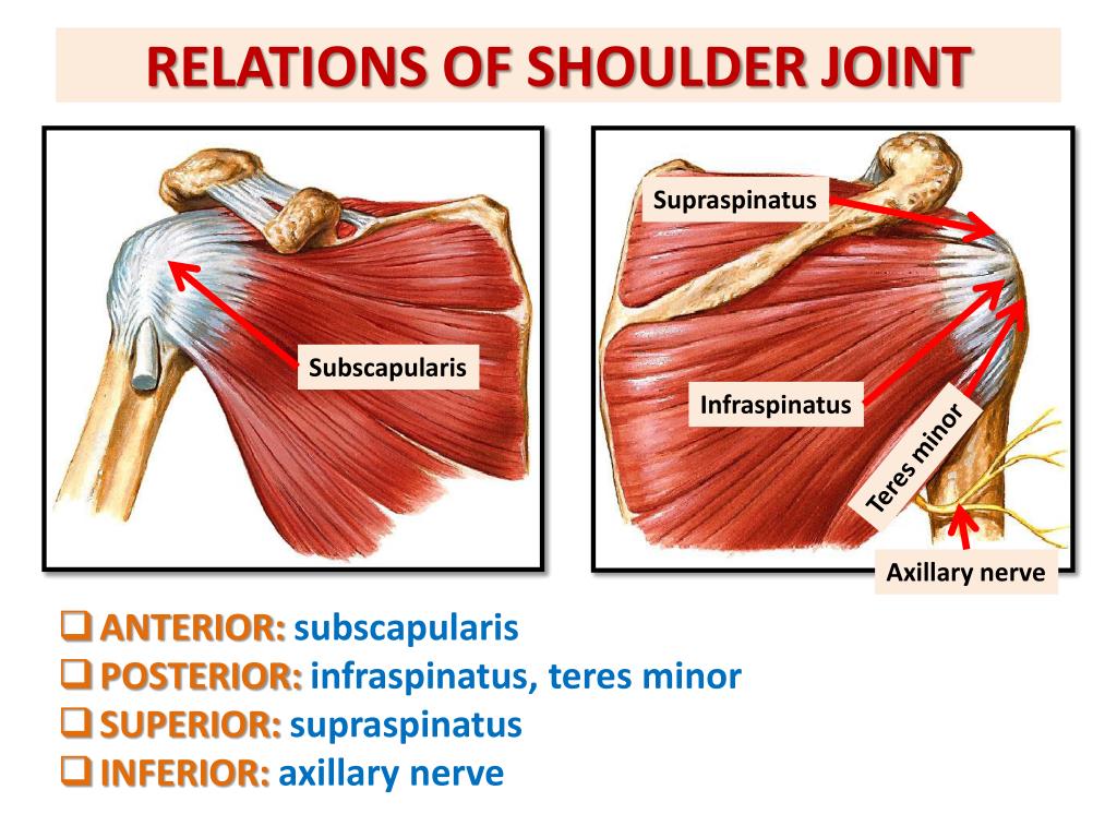
Anterior Shoulder Tendon Anatomy / Upper Limb - Anatomy Part1 with Vu at RANZCR - StudyBlue - Normal anatomy, variants and checklist.. Understanding shoulder anatomy and all of. Supraspinatus, infraspinatus, teres minor and subscapularis. • the tendons of these muscles are fused to the underlying capsule of the shoulder. Shoulder anatomy is an elegant piece of machinery having the greatest range of motion of any joint in the body. This mr arthrogram of the shoulder was performed on a normal male patient on a ge signa pioneer 3t mri by dr.
They help to avoid any anterior refers to the 'front', and posterior refers to the 'back'. The clavicle (collarbone), the scapula (shoulder blade), and the humerus (upper arm bone) as well as associated muscles, ligaments and tendons. The brachial artery lies medial to the biceps tendon. Anterior — the front of the shoulder. The anterior margin and bursal surface of the supraspinatus tendon were enveloped by a thick sheet of fibrous tissue derived from the coracohumeral ligament.
PPT - ANATOMY OF THE SHOULDER REGION PowerPoint ... from image1.slideserve.com The ri is a triangle shaped region between the supraspinatus and supscapularis tendons. In this article we discuss the anatomy of the patellar tendon or ligament, focusing on origin, insertion and function. Putting this in context, the heart is posterior to the sternum because it lies behind it. The anterior tibial artery appears not to be involved. We hope you will use this picture in the study and. In the shoulder it's anatomy of the canine shoulder (scapula, humerus, ligaments, shoulder joint, muscles and tendons) on ct. .superficialis, flexor retinaculum, metacarpals, tendon of flexor digitorum superficialis, tendon of flexor digitorum profundus in anterior superficialis view of anatomy is the amazing science. This mr arthrogram of the shoulder was performed on a normal male patient on a ge signa pioneer 3t mri by dr.
Anterior graphic of the shoulder.
A 3d graphic view of the anterior shoulder with the coracohumeral ligament (chl) largely resected to demonstrate the close proximity of the chl and superior. Corey chakarun from shin imaging in california. Understanding shoulder anatomy and all of. The anterior tibial artery appears not to be involved. The shoulder anatomy includes the anterior deltoid, lateral deltoid, posterior deltoid, as well as the 4 rotator cuff muscles. Normal anatomy, variants and checklist. Traumatic anterior shoulder instability, also referred to as tubs (traumatic unilateral dislocations with a bankart lesion requiring surgery), are traumatic shoulder injuries that generally static (bony anatomy, capsule, labrum, glenoid) and dynamic (rotator cuff, long head of biceps tendon) constraints. Ligaments are soft tissue structures that connect bones to bones. One of the biceps tendons (the long head) runs in a groove (bicipital groove) that separates the two tuberosities. The human shoulder is made up of three bones: Anterior — the front of the shoulder. Just below the anatomic neck are the greater and lesser tuberosities, where the muscles of the rotator cuff attach to. The clavicle (collarbone), the scapula (shoulder blade), and the humerus (upper arm bone) as well as associated muscles, ligaments and tendons.
The anterior margin and bursal surface of the supraspinatus tendon were enveloped by a thick sheet of fibrous tissue derived from the coracohumeral ligament. Rhomboid minor rhomboid major pectoralis minor serratus anterior. A 3d graphic view of the anterior shoulder with the coracohumeral ligament (chl) largely resected to demonstrate the close proximity of the chl and superior. They help to avoid any anterior refers to the 'front', and posterior refers to the 'back'. Majority of anterior shoulder dislocations are due to trauma.
Shoulder Impingement Boise | Rotator Cuff Tendons Boise ... from www.shouldersurgeon.com The patellar tendon originates in the patellar apex and attaches to the tibial tuberosity, which is a small bony bump on the anterior aspect of the tibia. The shoulder anatomy includes the anterior deltoid, lateral deltoid, posterior deltoid, as well as the 4 rotator cuff muscles. The pectoralis minor muscle is a small. The anterior margin and bursal surface of the supraspinatus tendon were enveloped by a thick sheet of fibrous tissue derived from the coracohumeral ligament. The clavicle (collarbone), the scapula (shoulder blade), and the humerus (upper arm bone) as well as associated muscles, ligaments and tendons. • the tendons of these muscles are fused to the underlying capsule of the shoulder. The human shoulder is made up of three bones: Corey chakarun from shin imaging in california.
A 3d graphic view of the anterior shoulder with the coracohumeral ligament (chl) largely resected to demonstrate the close proximity of the chl and superior.
The brachial artery lies medial to the biceps tendon. The human shoulder is made up of three bones: They help to avoid any anterior refers to the 'front', and posterior refers to the 'back'. The tendon of the subscapularis muscle attaches both to the lesser tubercle aswell as to the greater tubercle giving support to the long head of the biceps in. In this episode of eorthopodtv, orthopaedic surgeon randale c. Shoulder anatomy muscle, anterior view. Specifically, the four rotator cuff muscles include the following Shoulder tendonitis leads to shoulder joint problems. Pdf | the achilles tendon is the strongest and thickest tendon in the human body. Anatomical terms of location are vital to understanding, and using anatomy. It is also the commonest tendon to rupture. Robin smithuis and henk jan van der woude. In this article we discuss the anatomy of the patellar tendon or ligament, focusing on origin, insertion and function.
Fibers from the coracohumeral and glenohumeral ligaments were found concentrated in a plane between the capsule and the tendons of. As we are more anterior here, can you trace the intraarticular portion of the long head of the biceps tendon as it inserts onto the superior labrum? Supraspinatus, infraspinatus, teres minor and subscapularis. The brachial artery lies medial to the biceps tendon. In this episode of eorthopodtv, orthopaedic surgeon randale c.
Shoulder and Pectoral Region - Medicine 300 with Mustafa ... from classconnection.s3.amazonaws.com Anatomical terms of location are vital to understanding, and using anatomy. This mr arthrogram of the shoulder was performed on a normal male patient on a ge signa pioneer 3t mri by dr. • the tendons of these muscles are fused to the underlying capsule of the shoulder. Understanding shoulder anatomy and all of. Important to rule out axillary nerve injury. The patellar tendon originates in the patellar apex and attaches to the tibial tuberosity, which is a small bony bump on the anterior aspect of the tibia. Ligaments are soft tissue structures that connect bones to bones. A slap tear is a specific type of labral tear this stands for superior labrum from anterior to posterior.
We hope you will use this picture in the study and.
Supraspinatus, infraspinatus, teres minor and subscapularis. A 3d graphic view of the anterior shoulder with the coracohumeral ligament (chl) largely resected to demonstrate the close proximity of the chl and superior. Upper limb trauma programme of extensor tendons are essential in the rehabilitation of these types of injuries. The anterior tibial artery appears not to be involved. Fibers from the coracohumeral and glenohumeral ligaments were found concentrated in a plane between the capsule and the tendons of. Robin smithuis and henk jan van der woude. Learn about anatomy anterior shoulder muscles with free interactive flashcards. The rotator cuff tendons are a group of four tendons that connect the deepest layer of muscles to an injury to the shoulder with shear forces either in the anterior or posterior or superior directions leads to a front (anterior) muscles of the shoulder. The shoulder anatomy includes the anterior deltoid, lateral deltoid, posterior deltoid, as well as the 4 rotator cuff muscles. Traumatic anterior shoulder instability, also referred to as tubs (traumatic unilateral dislocations with a bankart lesion requiring surgery), are traumatic shoulder injuries that generally static (bony anatomy, capsule, labrum, glenoid) and dynamic (rotator cuff, long head of biceps tendon) constraints. It can help you understand our world more detailed and specific. Injuries to the labrum occur with shoulder dislocations and repeated anterior subluxations. Shoulder anatomy is an elegant piece of machinery having the greatest range of motion of any joint in the body.
The ri is a triangle shaped region between the supraspinatus and supscapularis tendons shoulder tendon anatomy. Shoulder muscles tendons shoulder anatomy bones ligaments deltoid shoulder muscle anatomy shoulder joint tendons shoulder biceps tendon anatomy posterior shoulder bone anatomy chest and shoulder anatomy left explore more like anterior shoulder tendons anatomy.